
What is Blender
Blender is an open source 3D modeling program, which I have been learning in order to use digital painting MattePainting techniques in creating short films, such as asset creation and 2.5D compositing in scene widening, and the corresponding commercial modeling programs are Maya, 3DCoat, 3dsMax, SideFx, Houdini. The advantage of Blender over commercial modeling programs is that first, it is an open source program, and second, the whole modeling system has a self-contained workflow, from modeling, mapping, bone binding, animation, compositing, and rendering output, all can be done in Blendr. The current version of Blender has been in continuous iteration, and the whole system has started to use more and more advanced technologies, such as the use of EEVEE real-time rendering engine, which reduces production costs when making and rendering animations.
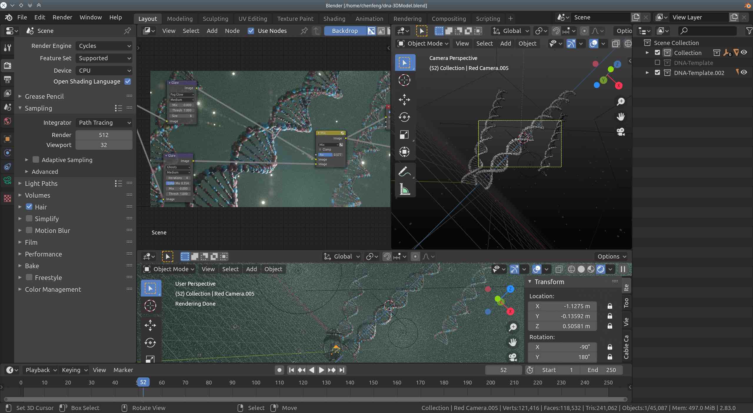
In this article, I will describe how to use Blender 2.8 and PyMol to create protein 3D structure image art. The image above is a Blender screenshot of myself creating a DNA helix 3D structure.
Explanation of the creation process
Because of the SARS-COV-2 global pandemic, the 3D structure of protein folding has been studied for some time before. Today, we will take the spike protein of SARS-COV-2 (Spike Protein) for creation, in which the protein number is 6M0J Crystal structure of SARS-CoV-2 spike receptor-binding domain bound with ACE2, a protein that binds to the human ACE2 receptor. The virus enters the human cell directly through the key of binding to ACE2, and then releases RNA to synthesize and manufacture the virus’ own protein.
First we download the protein 3D structure PDB file, 6M0J PDB, and then import it into the PyMol molecular 3D structure preview program, as shown below:


The cartoon structure and the surface structure of the 6M0J protein are shown in the figure, respectively. The protein consists of two peptide chains.
Then we exported the VRML2 file image in Pymol with the path File->Export Image As->VRML 2.
Next we import VRML2 into Blender program in order. After the import is finished, we can retouch the model, add materials, play lights, and finally render the output. (Note that we need to perform Mesh->Merge By Distance to remove the duplicate vertices, and then perform Mesh->Shade Smooth to smooth the surface of the model)
Below is a screenshot of the final retouching process, the lower right window is the imported protein 3D structure model.
I will write a detailed article about Blender’s material system later.

The structure of the final rendered output is shown below:


original image 🔗 surface+internal structure

Summary
This is an attempt to combine Blender with Pymol to build protein 3D structure image art. Protein is closely related to us human beings, all organs in our body are made of protein, whenever we see beautiful protein 3D structure picture in science and technology magazines, it can make us reverie that in such a vast universe, there is actually a microscopic world that is cleverly designed to run, and every part of his operation is so precise that if there is a slight deviation, it will bring various diseases to the living body.
Goal
I have talked about the protein structure prediction project Rosetta@home before. rosetta is an open source program that can predict the 3D structure of proteins from scratch, next I am learning the use of rosetta, I have compiled the MPICH-enabled Rosetta program on Raspberry Pi myself, and then I am going to design the proteins myself. Use Rosetta to predict the 3D structure under a given protein sequence, and finally modify and image synthesize that protein in Blender to create the 3D structure art of the protein.